Precise CLIP-seq Services
Home
> Precise CLIP-seq Services
Precise CLIP-seq Services – Map RNA-Protein Interactions for Epigenomics Research
N2 Jenomics Lab Pvt. Ltd. delivers research-grade CLIP-seq solutions to decode RNA–protein interactions at high resolution. Our expert team integrates advanced protocols and powerful bioinformatics to help researchers explore gene regulation, identify novel targets, and drive innovation in epigenomics.
Scientists often want to know how proteins interact with RNA inside cells. These interactions are important because they help control how genes work and how cells behave. CLIP-seq, which stands for "Crosslinking and Immunoprecipitation followed by Sequencing," is a method that helps researchers find out exactly where proteins bind to RNA.
In CLIP-seq, researchers shine ultraviolet (UV) light onto cells. This makes proteins and RNA stick together where they touch. Next, they use special antibodies to pull out these protein-RNA pairs from the rest of the cell. After that, they break the RNA into small pieces and turn it into DNA. Finally, they sequence this DNA to learn where the proteins were bound on the RNA.
Compared to a simpler method called RIP-seq (RNA Immunoprecipitation sequencing), CLIP-seq has two big advantages:
Higher specificity: UV light ensures that only proteins directly touching the RNA get linked, which reduces false positives.
Better resolution: CLIP-seq shows exactly which part of the RNA the protein was binding to, sometimes down to a few nucleotides.
CLIP-seq Technologies Packages
Classic CLIP-seq (HITS-CLIP)
Classic CLIP-seq, sometimes called HITS-CLIP, uses UV light to crosslink proteins and RNA directly inside cells. Scientists then use antibodies to pull out these protein-RNA pairs. Next, the RNA is converted into DNA and sequenced. This method provides a broad, genome-wide map of where proteins bind to RNA.
iCLIP-seq (Individual-nucleotide Resolution CLIP)
iCLIP-seq is designed to map protein–RNA interactions with single-nucleotide precision. During the reverse transcription step, iCLIP captures where the enzyme stalls due to crosslinked proteins, creating precise "cut-off" points.
This technology is particularly useful for studying:
Splicing regulation
RNA maturation
Non-coding RNA function
iCLIP-seq helps researchers identify exact protein binding positions on RNA, making it ideal for high-resolution studies.
miCLIP-seq is a specialized version of iCLIP used to study RNA modifications, particularly m6A methylation.
By combining UV crosslinking with specific antibodies against methylated bases, miCLIP-seq allows researchers to detect:
Single-nucleotide resolution methylation sites
Binding sites of "reader" proteins that recognize RNA modifications
This technique is crucial for understanding epitranscriptomic regulation and how chemical changes to RNA affect gene expression and cell function.
eCLIP-seq (Enhanced CLIP-seq)
eCLIP-seq improves on traditional CLIP methods by adding size-matched input controls to better distinguish real binding events from background noise.
Benefits of eCLIP-seq include:
Higher signal-to-noise ratio
Improved reproducibility
Greater confidence in identifying true protein-RNA interactions
eCLIP-seq has become a popular method for large-scale projects, such as ENCODE, where precise and reproducible data is essential.
PAR-CLIP-seq
PAR-CLIP-seq involves feeding cells molecules called photoreactive ribonucleoside analogs, like 4-thiouridine (4SU). These get incorporated into newly transcribed RNA. When UV light hits the cells, it promotes stronger crosslinking between the RNA and bound proteins.
A unique feature of PAR-CLIP is its ability to pinpoint exact binding sites at the nucleotide level, often detected through characteristic T-to-C conversions in the sequence data.
Comparison of ATAC-seq, ChIP-seq, CUT&Tag, RIP-seq, and CLIP-seq
Researchers often wonder which sequencing technology best fits their study. N2 Jenomics Lab Pvt. Ltd. offers all these methods to help you explore gene regulation from different angles. The table below makes it easy to compare these powerful tools:
Technology
Main Focus
Target Molecule
Resolution
Special Features
ATAC-seq
Finds open regions of DNA
DNA
High
Fast method to see chromatin accessibility
ChIP-seq
Maps protein binding on DNA
DNA
High
Great for studying transcription factors and histone modifications
CUT&Tag
Maps protein-DNA interactions with less background
DNA
Very High
Lower input requirements than ChIP-seq
RIP-seq
Studies protein-RNA interactions
RNA
Medium
Easier protocol but lower resolution than CLIP-seq
CLIP-seq
Pinpoints exact protein binding sites on RNA
RNA
Very High
UV crosslinking ensures high specificity and nucleotide-level resolution
Each method provides unique insights. You can choose a single technique or combine several for deeper analysis.
"At N2 Jenomics Lab Pvt. Ltd. , we are committed to staying one step ahead through constant innovation. We have built deep expertise in epigenomics, not only focusing on core technologies like methylation analysis, ATAC-seq, ChIP-seq, and RIP-seq, but also embracing cutting-edge methods such as CLIP-seq and CUT&Tag. Our goal is to help clients solve even the most complex challenges in epigenetic research."
CLIP-seq Experimental Workflow
CLIP-seq Bioinformatics
Core Data Processing
Once we receive the sequencing data, we start by cleaning it up. This involves:
Quality control: Checking for errors or low-quality reads that might cause problems later.
Adapter trimming: Removing leftover pieces of DNA used during library preparation.
Mapping reads: Aligning each RNA fragment to the right spot on the genome.
Advanced Analysis and Functional Interpretation
Peak calling: Identifying regions where many RNA fragments pile up, signaling a protein-binding site.
Motif discovery: Searching for short RNA sequences that proteins prefer to bind.
Positional analysis: Examining where binding sites occur, like near the start or end of genes (TSS, TTS, start/stop codons).
GO and KEGG pathway analysis: Linking bound genes to known biological functions and pathways.
Differential analysis: Comparing samples to see how binding changes under different conditions.
CIMS analysis: Detecting tiny changes in RNA caused by the crosslinking process, helping pinpoint binding sites.
We also create clear graphs and visualizations to help researchers see and interpret their results.
Multi-Omics Integration
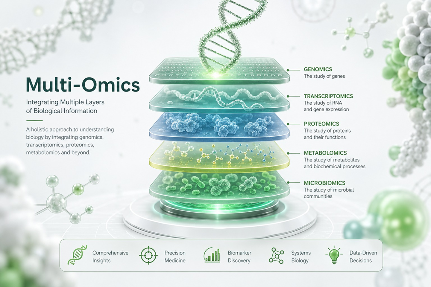
Modern research often involves multiple types of data. N2 Jenomics Lab Pvt. Ltd. helps researchers combine CLIP-seq with:
ChIP-seq: Studying protein-DNA interactions.
ATAC-seq: Finding open regions of DNA.
RNA-seq: Measuring gene expression levels.
Advantages of Our CLIP-seq Services
Extensive Experience Across Diverse Species and Sample Types
Our team has handled CLIP-seq projects for a wide variety of organisms, including:
Humans
Mice, rats, and other animals
Plants such as rice, wheat, and tomato
Microorganisms like bacteria and fungi
High Success Rates
CLIP-seq experiments can be complex. But at N2 Jenomics Lab Pvt. Ltd. , our success rates exceed 95%, thanks to:
Optimized lab protocols
Careful experimental design
Rigorous quality control
Proprietary Peak-Calling Algorithms
We've developed our own algorithms for peak calling, the critical step of identifying where proteins bind to RNA. Our proprietary methods:
Reduce background noise
Improve detection of real binding events
Deliver clearer, more reliable results
Customized Multi-Omics Analyses
Every research question is different. That's why we offer customized data analysis and multi-omics integration, allowing you to:
Connect CLIP-seq data with ChIP-seq, ATAC-seq, or RNA-seq results
Explore complex regulatory networks
Gain a deeper understanding of gene expression and regulation
Strict Antibody Quality Validation
Antibodies are the key tools in CLIP-seq. We:
Test every antibody for specificity and efficiency
Provide validation data to ensure confidence in results
Help clients choose the right reagents for their targets
Quick Turnaround Times and High-Quality Deliverables
We know that time matters in research. N2 Jenomics Lab Pvt. Ltd. offers:
Fast project turnaround
Detailed, publication-ready reports
Clean, well-organized data files for easy downstream analysis
Strong Publication Record
Our scientists have contributed to studies published in top-tier journals like Nature, Cell, and Molecular Cell. We bring that same level of scientific rigor to every client project.
Applications of CLIP-seq
1
Understanding RNA-Binding Proteins (RBPs)
How genes are turned on or off
How cells respond to stress
How different cell types develop unique identities
2
Studying Alternative Splicing
Discover new RNA variants
Understand how splicing errors contribute to disease
Identify new targets for drug development
3
Exploring Non-Coding RNAs (ncRNAs)
Discover how RBPs interact with non-coding RNAs
Reveal new functions for these mysterious molecules
Identify potential biomarkers for research applications
4
Identifying miRNA Targets
High-confidence identification of miRNA target sites
Insights into how miRNAs regulate specific biological pathways
5
Supporting Drug Target Discovery
Identify RBPs or RNA regions involved in disease-related pathways
Study how experimental drugs affect RNA-protein interactions
Find potential biomarkers for drug development
Sample Requirements
Our HLA typing service provides high-resolution data tailored to support diverse research applications. Here's a quick overview of how to interpret the results.
Sample Type
Recommended Quantity
Minimum Quantity
Minimum Concentration
Special Notes
Cells
≥ 1×10⁷ – 3×10⁷ cells
—
—
Number depends on target protein abundance and CLIP-seq method.
Tissue
≥ 10 – 500 mg
10 – 200 mg
—
Amount varies by tissue type and target protein. Consult N2 Jenomics Lab Pvt. Ltd. for specifics.
IPed RNA
≥ 100 ng
40 ng
≥ 5 ng/μL
Applicable if submitting purified RNA after immunoprecipitation.
Demo Results
Precise CLIP-seq FAQs
1. What is CLIP-seq and why is it important?
CLIP-seq combines UV crosslinking, immunoprecipitation, and sequencing to map RNA-protein interactions at a transcriptome-wide scale. This provides precise insights into how RNA-binding proteins regulate gene expression post-transcriptionally .
2. How does CLIP-seq differ from RIP-seq?
RIP‑seq pulls down RNA-protein complexes without crosslinking, leading to more background noise and less precise binding site identification. CLIP-seq uses UV crosslinking to capture direct, in vivo interactions, offering higher specificity and single-nucleotide resolution .
3. What resolution can I expect with CLIP-seq variants like iCLIP or PAR-CLIP?
iCLIP captures truncated cDNAs at crosslink sites, enabling individual-nucleotide resolution and precise protein–RNA binding location.
PAR-CLIP, using photoactivatable nucleosides, introduces characteristic mutations (e.g., T-to-C) that help pinpoint binding sites down to specific nucleotides.
Mapping reads to a reference genomePeak calling to identify binding sites
Motif discovery and binding site quality checks
Pipelines like CLIPipe, PureCLIP, and PEAKachu are commonly used for robust analysis workflows.
5. What types of samples and input quantities are needed?
CLIP-seq typically requires 10–30 million cells or 10–500 mg of tissue, depending on the specific method. Variants like GoldCLIP or irCLIP may accept lower inputs, but overall yield and protein abundance are critical considerations. It is best to consult CD Genomics for sample-specific advice.
6. Can CLIP-seq detect RNA modifications like m6A?
Yes. Variant methods such as miCLIP-seq combine CLIP with antibodies targeting modified RNA bases—especially m6A—to map modified nucleotides at single-nucleotide resolution, helping understand epitranscriptomic regulation.
7. How do I validate antibody specificity in CLIP-seq?
High-quality CLIP-seq relies on validated antibodies. Researchers should ensure their antibodies are specific and work well in immunoprecipitation. Tools like Western blot and IP-qPCR are recommended before proceeding to CLIP-seq.
8. What controls are necessary in CLIP-seq experiments?
Controls such as input samples, size-matched input (SMI) for eCLIP, and IgG or no-antibody controls are vital. These controls help distinguish true binding events from background noise.
9. How do I choose between CLIP variant methods?
Use iCLIP for nucleotide-precise mapping
Choose PAR-CLIP for strong signal via photoreactive nucleosides
Consider GoldCLIP or irCLIP for safer protocols without radioactivity
Use eCLIP for standardized input controls and robust reproducibility
Precise CLIP-seq Case Studies
Case Study: PRRC2B-mRNA Interactions Mapped by PAR-CLIP-seq
Journal: Nucleic Acids Research
Impact Factor: 19.160 (2022)
Published: 2023
PMC10287950
Background
RNA-binding proteins (RBPs) play crucial roles in regulating gene expression post-transcriptionally, influencing processes such as splicing, translation, and RNA stability. However, the functions and binding patterns of many RBPs remain poorly characterized. This study focused on PRRC2B, a poorly understood RBP suspected of influencing cell cycle progression and cancer-related pathways. The researchers aimed to identify genome-wide RNA binding sites of PRRC2B and explore its functional role in translational regulation.
Materials & Methods
Sample Preparation
Cell Line: HEK293T
Incorporation of photoreactive ribonucleoside analog 4-thiouridine (4SU)
UV crosslinking at 365 nm
Immunoprecipitation & Sequencing
Immunoprecipitation of PRRC2B-RNA complexes
Library preparation and PAR-CLIP sequencing
Data Analysis
Mapping reads to the human genome
Identification of crosslink-induced mutation sites (CIMS)
Motif analysis
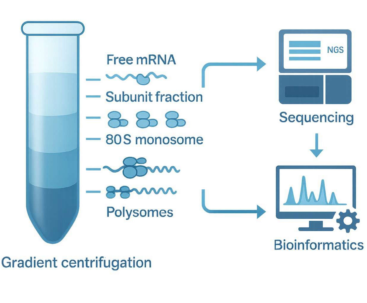
Ribosome profilingfor translation efficiency
Functional assays including PRRC2B knockdown and rescue experiments
Results
PRRC2B Binding Site Identification
Genome-wide binding sites of PRRC2B were mapped with single-nucleotide resolution.
PRRC2B preferentially bound near the start codon regions of a subset of mRNAs.
Enriched motifs included CU- or GA-rich sequences in 5' UTRs and coding sequences.
Functional Consequences of PRRC2B Binding
PRRC2B knockdown led to:
Reduced translation of target mRNAs
Decreased levels of cell-cycle regulatory proteins, including CCND2
G1/S phase arrest and reduced cell proliferation
Antisense oligonucleotides disrupting PRRC2B binding at specific motifs reduced translation of CCND2.
Mechanistic Insights
PRRC2B interacts with translation initiation factors eIF3 and eIF4G2 in an RNA-independent manner.
Mutations disrupting PRRC2B's interactions with initiation factors failed to rescue translation defects caused by PRRC2B knockdown.
Proline-rich coiled-coil 2B (PRRC2B) binds to CU- or GA-rich motifs on specific mRNAs, facilitates their translation through interacting with eIF4G2 and eIF3, and promotes cell cycle progression and cell proliferation.
Conclusion
This study provides the first comprehensive, high-resolution map of PRRC2B-RNA interactions in human cells, demonstrating that PRRC2B binds near translation start sites and enhances translation of key cell-cycle regulators. Disruption of these interactions impairs cell cycle progression, suggesting a novel mechanism for translational control. These insights highlight PRRC2B as a potential regulatory node in cancer biology and cell proliferation pathways.
Reference
Jiang, F., Hedaya, O. M., Khor, E. S., et al. (2023). RNA binding protein PRRC2B mediates translation of specific mRNAs and regulates cell cycle progression. Nucleic Acids Research. PMC10287950
Address:Registered Office: 150, Patparganj Industrial Area, New Delhi – 110092, India
Operational Address:National Institute of Plant Genome Research (BRIC - NGGF)
Lab No. 206 and 207, Aruna Asaf Ali Marg, P.O. Box No. 10531,
New Delhi – 110067, India