Multiplex Ligation Dependent Probe Amplification Assay Service: Precision CNVs Analysis Made Simple with MLPA
N2 Jenomics Lab Pvt. Ltd. MLPA (Multiplex Ligation-Dependent Probe Amplification) service offers a quick and accurate way to detect DNA copy number variations (CNVs) linked to genetic disorders and cancer. MLPA is effective for detecting CNVs, especially in cases with complex gene sequences, and is commonly used as a first-line test. Using a simple, multiplex PCR-based method, MLPA amplifies probes with unique genomic targets, which are fluorescently labeled and quantified through capillary electrophoresis. This allows for precise analysis of genetic variations, making it a powerful tool for both research and clinical applications.
What is Multiplex Ligation-Dependent Probe Amplification (MLPA)?
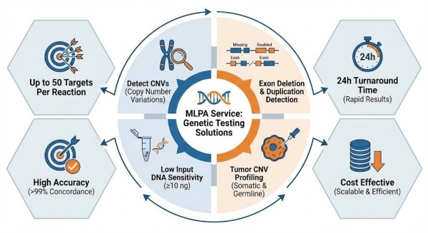
Multiplex Ligation-Dependent Probe Amplification (MLPA) is a widely used molecular biology technique for detecting the copy number of various DNA sequences in human genetic disease research. This technique involves the ligation of probe oligonucleotides followed by PCR amplification, allowing the analysis of up to 50 multiplex probe pairs designed to hybridize with specific target loci.
Each probe pair is designed to generate amplification products of a specific length. By incorporating universal sequences at their termini, all ligated probes can be amplified in a single PCR reaction using one primer pair. The forward PCR primer is labeled with an Applied Biosystems™ 6-FAM™ fluorescent tag, enabling detection and quantification based on the molecular sizes of the probes, as determined through automated capillary electrophoresis (CE).
Multiplex Ligation-Dependent Probe Amplification (MLPA) process visualized in three steps: 1) Denaturation & Hybridization, 2) Ligation, 3) Amplification with fluorescent PCR primers to detect copy number variations.
This method has been successfully used in the study of diseases caused by exon deletions and duplications, such as Duchenne Muscular Dystrophy (DMD) and BRCA1/BRCA2 gene mutations. Additionally, advancements in the MLPA technique now enable its application in quantitative methylation analysis of various genomic sequences.
Why Choose MLPA?
MLPA is preferred for its precise, high-throughput analysis, even with limited DNA samples. Unlike traditional PCR, MLPA can test multiple genetic loci at once, making it a cost-effective and reliable solution for detecting genetic abnormalities. It is also more sensitive than other multiplex PCR methods, offering better detection of copy number variations (CNVs).
MLPA combines DNA probe hybridization with PCR technology, offering the following advantages:
High Efficiency: A single reaction can detect copy number variations in up to 50 target sequences.
Rapid Turnaround: Complete experiments can be performed within 24 hours.
Operational Simplicity: Different reagent kits adhere to nearly identical procedures, rendering the technique easy to learn and master.
Technical Comparison: MLPA and Alternative Genetic Analysis Methods
Parameter
MLPA
qPCR
ddPCR
Sanger Seq
Targeted NGS
WES
WGS
CGH Microarray
Multiplex Level
Up to 50 targets
Few
Few
N/A
Dozens–Hundreds
Thousands
Whole genome
Genome-wide
Main Purpose
CNV detection
Quantitation
Quantitation
Sequence variant validation
Variant detection (panel)
Exon variants
All variants & SV
CNV genome-wide
Sensitivity for CNV
High
Moderate
Very high
Low
High
Medium
High
High
Point mutation detection
No
Rare
Limited
Yes
Yes
Yes
Yes
No
Equipment Needed
PCR + CE
qPCR machine
ddPCR instrument
Sanger sequencer
NGS instrument
NGS
NGS
Microarray scanner
Throughput
Medium
Low
Low
Low
Medium
Medium
High
High
Quantitative Accuracy
Good
Variable
Excellent
n/a
Good
Good
Good
Good
Turnaround time
~1 day
~1 day
~1 day
~1 week
~1–2 weeks
~2–4 weeks
~3–6 weeks
~2–3 weeks
Cost
Low
Low
Medium
Medium
Medium
High
Highest
High
Applications of MLPA
MLPA is a trusted method for detecting genetic variations with high sensitivity, and it finds application across various research and clinical fields. It enables researchers to analyze structural variations in the genome, providing insights into genetic disorders and disease mechanisms. This versatile technology supports a wide range of scientific inquiries, including but not limited to:
Detection of Small-Scale Gene Rearrangements:
Validated genes include BRCA1, BRCA2, MSH2, MLH1, DMD, APC, SMA, NF1, NF2, VHL, TSC1/2 etc.
Detection of Large-Scale Genomic Rearrangements:
Validated disorders include Williams syndrome, Prader-Willi/Angelman syndrome, DiGeorge syndrome, Cri du Chat syndrome, Pelizaeus-Merzbacher disease, CMT1A, and HNPP.
Sub telomeric Copy Number Variations
Chromosomal Aneuploidy Testing
Tumor Diagnosis Research:
DNA copy number analysis in malignancies such as acute lymphoblastic leukemia (ALL), chronic lymphocytic leukemia (CLL), oligodendrogliomas, melanomas, and neuroblastomas.
Quantitative Methylation Analysis:
Applications include Prader-Willi/Angelman syndrome, Beckwith-Wiedemann syndrome, MGMT, MLH1, Fragile X syndrome, and tumor suppressor gene inactivation.
mRNA Expression Analysis:
Focusing on genes involved in apoptosis and inflammatory response.
Multiplex Ligation Dependent Probe Amplification Assay Service Workflow
At N2 Jenomics Lab Pvt. Ltd. , we provide a streamlined, end-to-end MLPA service to ensure consistent, high-quality results. Our standardized workflow is designed to support reproducibility and accelerate discovery across genomic studies.
1. Sample Submission
Submit your DNA samples, including tissue, blood, or cell lines. Our team ensures that they meet quality standards for analysis.
2. DNA Extraction and QC
If needed, we offer DNA extraction services and perform quality checks to ensure optimal DNA quality for MLPA.
3. Probe Hybridization and Ligation
Probes are hybridized to your DNA and ligated to target regions for CNV analysis.
4. PCR Amplification
Multiple DNA regions are amplified simultaneously using fluorescent primers, enabling efficient CNV detection.
5. Capillary Electrophoresis
The amplified products are analyzed by capillary electrophoresis for accurate CNV quantification.
6. Data Analysis and Reporting
We provide detailed statistical and annotation reports, along with graphical results for easy interpretation.
Ship samples on blue ice or dry ice to preserve integrity.
DNA extraction services available upon request.
For special sample types or low-input scenarios, contact us for a customized plan.
Why Choose N2 Jenomics Lab Pvt. Ltd. for MLPA Service?
N2 Jenomics Lab Pvt. Ltd. offers a comprehensive MLPA service with expert support, fast results, and reliable data. Our team ensures high-quality testing using the latest technology, with quick turnaround times and global support. We follow strict quality control to provide accurate and dependable results for your research needs.
Expert Team: Our team of geneticists and molecular biologists ensures high-quality results with every test.
State-of-the-art Technology: We use the latest MLPA platforms to guarantee accurate and reliable data.
Fast Turnaround: Receive your comprehensive MLPA report in a timely manner, ensuring that your research or clinical decision-making isn't delayed.
Global Reach: Serving clients across the globe with customer support in multiple time zones.
Quality Assurance: We follow rigorous quality control protocols, ensuring that our tests meet the highest standards of accuracy and reliability.
References:
de Boer, S., White, S.J. Genotyping multiallelic copy number variation with multiplex ligation-dependent probe amplification (MLPA). In: Genotyping. Springer, New York, NY, 147–153 (2017). https://doi.org/10.1007/978-1-4939-6442-0_9
Cuevas, D., Velasco, A. et al. Intratumor heterogeneity in endometrial serous carcinoma assessed by targeted sequencing and multiplex ligation-dependent probe amplification (MLPA): a descriptive study. Histopathology 75, 724–733 (2019). DOI: 10.1111/his.14001
Fu, X., Shi, Y., Ma, J. et al. Advances of multiplex ligation-dependent probe amplification technology in molecular diagnostics. BioTechniques73, 205–213 (2022). DOI: 10.2144/btn-2022-0017
Demo Results
Figure 1. MLPA electropherogram comparison showing reference control peaks and reduced peaks in the test sample, indicating copy number deletions in specific gene loci.
Figure 2. Infographic MLPA result showing comparative bar peaks between control and test sample across multiple gene loci, with highlighted copy number variation.
Figure 3. MLPA result visualization in software UI style, displaying reference vs sample plots with highlighted copy number variation signals.
MLPA Assay FAQs
1. What types of samples can be used for MLPA?
You can use genomic DNA extracted from various sources such as blood, saliva, or tissue samples.
2. Can MLPA detect point mutations?
MLPA is primarily used for detecting copy number variations (deletions, duplications), but it can be adapted for mutation detection in specific cases.
3. What is the cost of MLPA testing?
The cost of MLPA testing varies depending on the number of targets analyzed. Please contact us for a quote.
MLPA Assay Case Studies
Reference
Xia Y., Feng Y., Xu L., Chen X., Gao F., Mao S. (2021). Case Report: Whole-Exome Sequencing With MLPA Revealed Variants in Two Genes in a Patient With Combined Manifestations of Spinal Muscular Atrophy and Duchenne Muscular Dystrophy. Frontiers in Genetics, Volume 12, Article 605611. DOI:10.3389/fgene.2021.605611.
1. Background
This case involves an 11-month-old male patient presenting with poor motor development and progressive muscle weakness. Clinical features resembled both Spinal Muscular Atrophy (SMA) and Duchenne Muscular Dystrophy (DMD), which are distinct neuromuscular genetic disorders. Accurate genetic diagnosis was crucial given overlapping symptoms and implications for treatment.
2. Methods
To pinpoint the genetic cause(s), clinicians used a two-tier genetic testing approach:
Whole-Exome Sequencing (WES) to screen for sequence variants across all coding regions.
Multiplex Ligation-Dependent Probe Amplification (MLPA) to specifically detect copy number variations (CNVs) such as deletions in target genes.
MLPA is a well-established method for exon-level CNV detection, making it suitable for DMD and SMA loci.
3. Results
The combined analysis identified:
A homozygous deletion of exons 7 and 8 in theSMN1gene, consistent with SMA.
A deletion in exon 50 of theDMDgene, diagnostic of DMD.
These findings were confirmed by MLPA following WES predictions.
MLPA genetic test reports showing zero copy of SMN1 exons 7 and 8 and a homozygous deletion of DMD exon 50 in the patient sample, compared to reference controls.
4. Conclusions
The integration of MLPA and WES enhanced diagnostic accuracy in this complex case involving dual neuromuscular disorders. This approach highlights the value of combining broad variant detection (WES) with targeted CNV profiling (MLPA) for comprehensive genetic diagnosis in clinically overlapping phenotypes.
Address:Registered Office: 150, Patparganj Industrial Area, New Delhi – 110092, India
Operational Address:National Institute of Plant Genome Research (BRIC - NGGF)
Lab No. 206 and 207, Aruna Asaf Ali Marg, P.O. Box No. 10531,
New Delhi – 110067, India